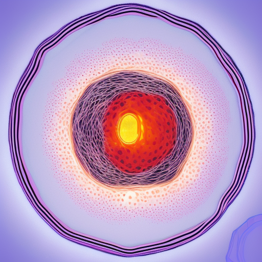
All cells possess a surrounding envelope known as the cell membrane, or plasma membrane. Animal cells are considered eukaryotic, meaning they have a well-developed nucleus and other membrane-bounded organelles that perform specialized functions. Figure 1.1 provides an illustration of a typical eukaryotic cell and some of its organelles. The cytoplasm is the part of the cell outside the nucleus and bounded by the cell membrane. The cytosol, or intracellular fluid, is the liquid portion of the cytoplasm, exclusive of organelles, and comprises a complex mixture of substances dissolved or suspended in water. The cell membrane is extensively discussed in Sections 2.1 and 2.2. The following cell organelles are of particular relevance for our purposes.
The cell’s endoplasmic reticulum (ER) is a complex network of vesicles, tubules, and cisternae. The rough endoplasmic reticulum (RER) is characterized by ribosomes, which are granular structures responsible for protein synthesis. These ribosomes can also be found free in the cytosol, as well as membrane-bound in the RER. Therefore, the RER is involved in synthesizing integral membrane proteins and proteins that are secreted outside the cell, such as hormones. On the other hand, the smooth endoplasmic reticulum (SER) is mostly without ribosomes and functions in lipid and steroid synthesis, as well as Ca2+ homeostasis by storing the ion in the cell. Muscle cells contain a type of SER, the sarcoplasmic reticulum (SR), which is specialized in taking up and releasing Ca2+.
The mitochondrion, as shown in Figure 1.2, typically has an elongated morphology with a diameter ranging from 0.1 to 0.5 µm and a length of one to several micrometers, though occasionally they can reach up to 20 µm in length. The number of mitochondria present in a cell varies significantly among different tissues and organisms, ranging from a single mitochondrion to several thousand per cell. Like the nucleus, the mitochondrion is enclosed by a double membrane with the inner membrane forming folds, known as cristae, which considerably increases the inner membrane’s surface area. Mitochondria can undergo fission and fusion independently of the cell to fulfill the cell’s specific demands. Additionally, they possess their own DNA (mtDNA) that codes for mitochondrial RNAs and proteins. mtDNA is inherited only from the mother and changes gradually over time. Despite having multiple vital functions, including participation in cell death, two functions are particularly important for our objectives:
Mitochondria serve as the cell’s primary source of adenosine triphosphate (ATP), which powers energy-consuming activities such as ionic pumps and muscular contraction. The hydrolysis of ATP to adenosine diphosphate (ADP) releases energy that drives various cellular processes. Phosphorylation, the addition of a phosphate group to a protein, is often used to activate proteins for action. ATP donates a phosphate group to the protein, becoming converted to ADP in the process. In later chapters, we will explore numerous examples of proteins being activated through phosphorylation and deactivated by dephosphorylation.
The mitochondrion has a strong attraction to Ca2+, which it stores temporarily, leading to a significant interplay of Ca2+ between the mitochondria and the SER. Ca2+ is taken up into the matrix, which is enclosed by the inner membrane, by a highly efficient Ca2+ uniporter (Section 2.3.2), and its concentration is practically the same as in the cytosol. Ca2+ is extruded by an antiporter (Section 2.3.2) in the inner mitochondrial membrane as well as by a complex channel called the mitochondrial permeability transition pore (mPTP).

Leave a Reply